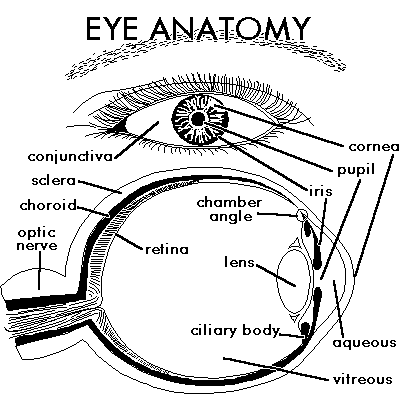
Eye Anatomy
Internal and External Structures of the Eye

Glossary of Eye Anatomy
- Aqueous
- A water-like fluid which fills the front part of the eye between the lens and cornea. This fluid is produced by the ciliary body and drains back into the blood circulation through channels in the chamber angle. It is turned over every 100 minutes.
- Chamber Angle
- Located at the junction of the cornea, iris, and sclera, the anterior chamber angle extends 360 degrees at the perimeter of the iris. Channels here allow aqueous fluid to drain back into the blood circulation from the eye. May be blocked in glaucoma.
- Choroid
- A very vascular layer between the sclera and retina which serves to nourish the outer portions of the retina. Has one of the highest blood flows in the body.
- Ciliary Body
- A structure located behind the iris (rarely visible) which produces aqueous fluid that fills the front part of the eye and thus maintains the eye pressure. It also allows focusing of the lens.
- Conjunctiva
- A thin lining over the sclera, or white part of the eye. This also lines the inside of the eyelids. Cell in the conjunctiva produce mucous, which helps to lubricate the eye.
- Cornea
- The clear window through which we see. Actually, this is a very vital part of the eye's focusing, and the curvature of the cornea itself accomplishes about 80% of the focusing of the eye.
- Episclera
- A fibrous layer between the conjunctiva and sclera. Sometimes lumps (pingueculum) will form in this layer on the surface of the eye near the inside or outside corners.
- Extraocular Muscles
- Six muscles control eye movement. Five of these originate from the back of the orbit and wrap around the eye to attach within millimeters of the cornea. Four of these move the eye roughly up, down, left and right. Two muscles (one originating from the lower rim of the orbit) control the twisting motion of the eye (when the head is tilted).
- Iris
- This is the part of the eye which gives it color. It contains muscles which open or close the pupil in response to the brightness of surrounding light. A blue iris actually has a lack of pigment.
- Lens
- This is located just behind the iris, and helps to focus light. A "capsule" surrounds the lens "nucleus". The nucleus can become cloudy, and this is termed cataract.
- Macula
- The part of the retina which is most sensitive, and is responsible for the central (or reading) vision. It is located near the optic nerve directly at the back of the eye (on the inside). This area is also responsible for color vision.
- Optic Nerve
- This contains visual information from the eye, and has about 1.2 million nerve fibers. The optic disc is visible on the inside of the eye, where the nerve is viewed "end on". The sheath around the optic nerve is continuous with that of the brain, and the nerve connects directly into the brain.
- Orbit
- The boney socket containing the eye, fat, extraocular muscles, nerves, and blood vessels. The floor and inside walls of the orbit are paper thin, and are easily fractured by trauma.
- Pupil
- Essentially, a hole in the iris. This is the black opening in the center of the eye. Its size is controlled by the iris muscles.
- Retina
- This thin layer lines the inside of the eye and receives light rays, processes them, and sends signals to the brain via the optic nerve. The retina is like the "film of a camera". It is separated from the very vascular choroid by the "retinal pigment epithelium". Sometimes breakdowns in this pigmented layer allow macular degeneration.
- Sclera
- The white, tough wall of the eye. Few diseases affect this layer. It is covered by the episclera and conjunctiva, and eye muscles are connected to this.
- Uveal Tract
- A group of similar eye structures including the choroid, ciliary body and iris. May be prone to inflammatory conditions (uveitis or iritis).
- Vitreous
- A jelly-like, clear fluid which fills most of the eye (from the lens back). This tends to liquefy with age, and its separation from the retina can lead to retinal tears and detachment.


